Let me start with a confession: the first time I ever saw a surgical microscope, I got vertigo. There’s nothing remotely ordinary about brain surgery, and when I first stepped into an operating room where every second counts, I realized the gravity of the moment—especially so when a patient younger than myself was wheeled in with a daunting diagnosis: glioblastoma. It wasn’t just about science, it was personal, complicated, and honestly, a little terrifying. This is a peek behind the blue drapes, beyond the clinical notes—into the unpredictable world where surgery, technology, and human resilience collide.
When Headaches Speak Louder: Recognizing the Unseen Signs
It’s easy to dismiss a nagging headache or a bit of clumsiness as stress or lack of sleep—especially in young adults. But sometimes, these symptoms are the body’s way of speaking up about something far more serious. I remember a night shift when a patient in their early 20s arrived with a story that started just like this: weeks of headaches, a limp that friends thought was just fatigue, and a growing sense of imbalance. It wasn’t until the left-sided weakness became impossible to ignore that the real story began to unfold.
Why a Lingering Headache and Shaky Balance Can Signal More Than Stress
When a headache persists for weeks, especially if it’s paired with new trouble walking or weakness on one side of the body, it’s time to look deeper. In this case, the patient’s symptoms included:
- Progressive headache lasting several weeks
- Left-sided hemiparesis (weakness on one side)
- Difficulty with gait and balance
On physical exam, the patient was alert and oriented. But subtle clues—like trouble moving the left arm and leg—hinted at something affecting the brain’s motor pathways.
Corticospinal Tract Dysfunction Symptoms: More Than Meets the Eye
The corticospinal tract is the main highway for voluntary movement, running from the brain down the spinal cord. When this pathway is disrupted, it can cause weakness, clumsiness, and abnormal reflexes. In this patient, two classic signs stood out:
- Hoffman Sign: Flicking the patient’s finger caused an involuntary twitch in the thumb and index finger. This is known as a positive Hoffman sign, named after German neurologist Dr. Johann Hoffman. It suggests dysfunction in the corticospinal tract, usually above the cervical spinal cord.
- Babinski Reflex: Stroking the sole of the foot made the big toe move upward—a positive Babinski sign. As I often explain,
‘A positive Babinski… is named after French neurologist Joseph Babinski.’
This reflex is a red flag for corticospinal tract disruption anywhere along its course.
Both signs are crucial bedside clues for upper motor neuron lesions. Their presence means the problem is not just in the muscles or nerves, but higher up—often in the brain or spinal cord.
Hoffman Babinski Reflex Significance: Why Those Odd Twitches Matter
In the operating room, these reflexes guide us to the root of the problem. A positive Hoffman or Babinski isn’t just a curiosity—it’s a signal that the brain’s control center is under threat. For this patient, the combination of symptoms and reflexes pointed to a lesion affecting the right side of the brain, which controls the left side of the body.
MRI Brain Tumor Diagnosis: The Critical Next Step
When neurological signs like these appear, an MRI brain tumor diagnosis becomes the frontline tool. In this case, the scan revealed a large mass at the temporal-parietal junction—confirming what the reflexes had already hinted. For young adults, especially, MRI is essential to catch these hidden threats early and plan the next steps in care.
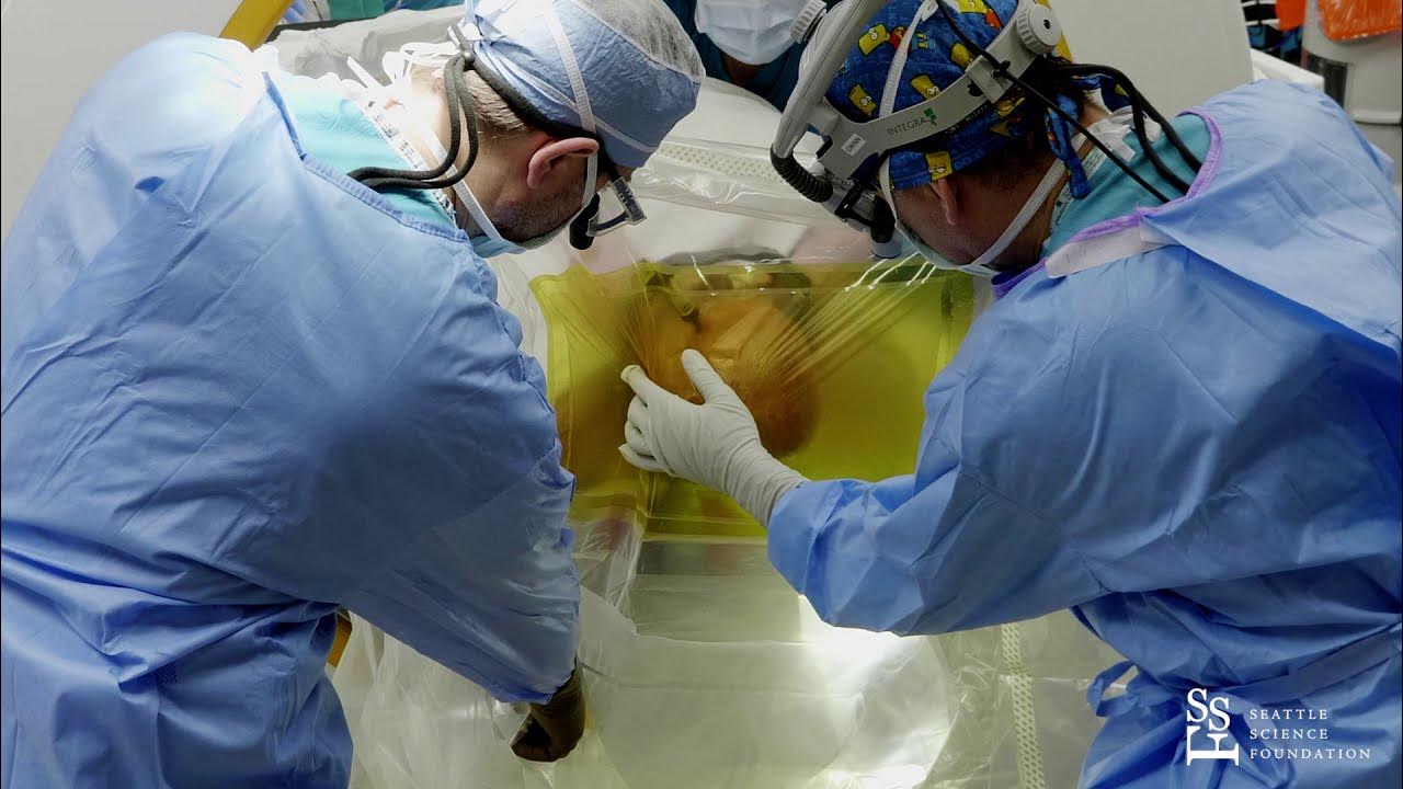
Blueprints, Gadgets, and Guts: Surgical Innovation in Real Time
Neurosurgery tumor resection today is a blend of careful planning, high-tech gadgets, and the willingness to adapt mid-procedure. When I step into the operating room for a glioblastoma case, it feels like entering a sci-fi lab—computer navigation neurosurgery, intraoperative brain mapping, and sodium fluorescent-guided microscopy are all part of the toolkit. But as my mentor used to say, “Everyone has a plan until we see the dura.” That’s when the real work—and the surprises—begin.
Patient Positioning: The First Blueprint
Patient positioning in neurosurgery is more than just comfort. For this case, the patient was placed supine, with a shoulder roll to elevate the right shoulder and the head gently turned left. This exposed the temporal parietal area, giving us the best corridor to the tumor. A single degree too far, and we risked damaging eloquent cortex—sometimes the difference between speech and silence post-op. Every angle matters.
Computer Navigation and Intraoperative Brain Mapping
Before the first incision, computer navigation neurosurgery helped us localize the tumor and plan a precise trajectory. We mapped the incision to bisect the tumor’s anterior and posterior poles, using advanced imaging to minimize risk to critical brain structures. Once the bone flap was elevated and the dura relaxed with hypertonic saline and mannitol, we could see the tumor clearly against normal brain tissue.
Intraoperative brain mapping and neurophysiological monitoring are our guides. We delivered electrical stimuli to the exposed cortex, starting at 2 milliamps and increasing in increments, to identify the primary sensory cortex and avoid it during resection. This real-time feedback is essential—no static MRI can match the detail we get from the living brain.
Surgical Techniques and the Role of Gadgets
With the surgical corridor optimized, we used bipolar cautery and microsuction to define the border between tumor and brain. Sometimes, a low-current bipolar cutter was needed to harden the tumor capsule, making it easier to distinguish from delicate brain tissue. These surgical techniques for brain tumors are refined, but the real leap comes from our next gadget: sodium fluorescent-guided microscopy.
Sodium Fluorescent-Guided Microscopy: Seeing the Unseen
Here’s where science fiction meets reality. Sodium fluorescent-guided microscopy highlights tumor cells that even the best eyes might miss. After what looked like a gross total resection, I was haunted by the possibility of hidden tumor. As I scanned the surgical field, the fluorescent dye revealed a suspicious area in a blind corner—
“Sodium fluorescent-guided microscopy showed an area in the blind corner suspicious for residual tumor.”
—and sure enough, additional tumor was found and safely removed.
Unexpected Lessons and Surgical Guts
Even with all the blueprints and gadgets, surgical guts matter. Plans change with the patient’s anatomy. The real fear in neurosurgery tumor resection isn’t what you see—it’s what you might leave behind. Sodium fluorescent-guided microscopy made me more vigilant, almost paranoid, about hidden corners. It’s a reminder that in brain surgery, innovation is as much about humility as it is about technology.
Hemming in the Bleeders: Old-School and New-School Hemostasis
When it comes to hemostasis techniques in brain surgery, the goal is simple: keep blood loss minimal and the surgical field clear. But the methods we use are a blend of time-tested tradition and cutting-edge science. In the operating room, the invisible labor of hemostasis is what lets us close up safely—and if we do it right, most patients and readers will never even notice this crucial step.
Why Hemostasis Matters in Brain Tumor Surgery
Bleeding in brain surgery isn’t just a nuisance—it can be life-threatening. Even a small amount of blood in the wrong place can cause pressure, swelling, or damage to delicate brain tissue. Effective surgical resection techniques depend on keeping the field dry and the patient safe. That’s why hemostasis is woven into every step of advanced surgical techniques for brain tumors.
Old-School: Hydrogen Peroxide and Cotton Balls
Some techniques have been around for decades, and for good reason. After removing a glioblastoma, I often reach for a time-honored tool: hydrogen peroxide solution. As the source notes:
‘Hydrogen peroxide solution soaked cotton balls is laid over the cavity… for both destruction of cells… and also for hemostasis.’
Hydrogen peroxide does double duty. It helps destroy any stray tumor cells that might remain in the cavity, and it also helps stop bleeding. The fizz and gentle pressure from the soaked cotton balls help seal off tiny blood vessels. More than once, the familiar smell of peroxide in the air has told me we’re nearing the end of a long case—a full circle moment that always brings relief.
New-School: Modern Hemostatic Agents
But we don’t stop at peroxide. Today’s hemostasis techniques in brain surgery include a range of advanced agents. Once the cavity is dry, we often cover it with specialized hemostatic materials—powders, gels, or sponges that promote clotting and seal off any oozing vessels. These agents are designed to work quickly and safely, reducing the risk of post-operative bleeding.
- Hydrogen peroxide: For both cell destruction and initial bleeding control
- Hemostatic agents: Applied to the cavity for rapid, reliable hemostasis
- Physical techniques: Gentle pressure, irrigation, and careful cautery
Closing Up: The Final Steps
Once we’re confident the bleeding is controlled, we move on to closure. The dura (the brain’s protective covering) is reapproximated and reinforced—a process called duroplasty. The bone flap goes back in place, and the border between the bone and the skull is sealed with a synthetic agent for a better cosmetic result. Each step relies on the success of the hemostasis that came before.
In the end, the art of hemostasis is a quiet but essential part of every brain tumor surgery. It’s a mix of old-school wisdom and new-school innovation, all working together to give patients the safest outcome possible.
Beyond the Surgery: Results, Surprises, and Human Realities
There are moments in the operating room that even the most experienced surgeons never forget. For me, one of those moments came when this patient, after a long and complex glioblastoma multiforme surgery, woke up with no new neurological deficits. Watching a patient walk out of the hospital, fully intact, is a result that stuns even the most jaded among us. Yet, as we celebrated this immediate post-operative outcome, the reality of the diagnosis set in: glioblastoma multiforme, an aggressive brain tumor with a reputation for returning, no matter how clean the initial resection appears.
The post-operative glioblastoma outcomes in this case were as good as anyone could hope for. The patient was discharged home, neurologically intact. The first MRI after surgery showed no evidence of residual tumor. Even more encouraging, the follow-up MRI a year later was also clear. As I reviewed the scans, I found myself repeating the words:
‘Post-operative MRI scan of the brain immediately and approximately a year after showed no radiographic evidence of residual or recurrent tumor.’
It’s the kind of result we all hope for, but rarely see in the world of glioblastoma multiforme surgery.
But optimism in glioblastoma care is always balanced by caution. The pathology report told a more complicated story. The tumor was confirmed as glioblastoma multiforme, with MGMT promoter methylation—a genetic marker that can predict better response to certain chemotherapies. However, there was no IDH1 or IDH2 mutation detected, which is associated with a less favorable prognosis. These genetic details matter. They shape our conversations about long-term follow-up glioblastoma treatment and help guide future therapy options. Even after a maximal safe resection, the risk of recurrence is high, and ongoing surveillance is not just recommended—it’s essential.
Managing tumor resection complications is always a concern, but in this case, the absence of new deficits was a testament to the precision of modern surgical techniques. Still, the human reality is that glioblastoma is relentless. We walk a fine line between the hope that comes with a clean MRI and the sobering statistics that define this disease. Every scan, every follow-up visit, is a reminder of both the progress we’ve made and the challenges that remain.
Sometimes, I find myself wondering about the future. What if artificial intelligence could one day spot every last tumor cell, even those invisible to the human eye? Maybe then, the art and science of glioblastoma surgery would tip further in our favor. For now, though, it remains a blend of technology, skill, and a lot of follow-up. The journey doesn’t end in the operating room—it continues with each scan, each conversation, and each moment of hope.
In the end, this patient’s story is a reminder of what’s possible, but also of the human realities we face every day. Glioblastoma multiforme surgery can deliver remarkable results, but the need for vigilance never fades. As we look to the future, we hold onto both the surprises and the hard truths, always striving for better outcomes in a field where every victory counts.
Leave a Reply