Introduction
In this lecture we re gonna cover the pharmacology
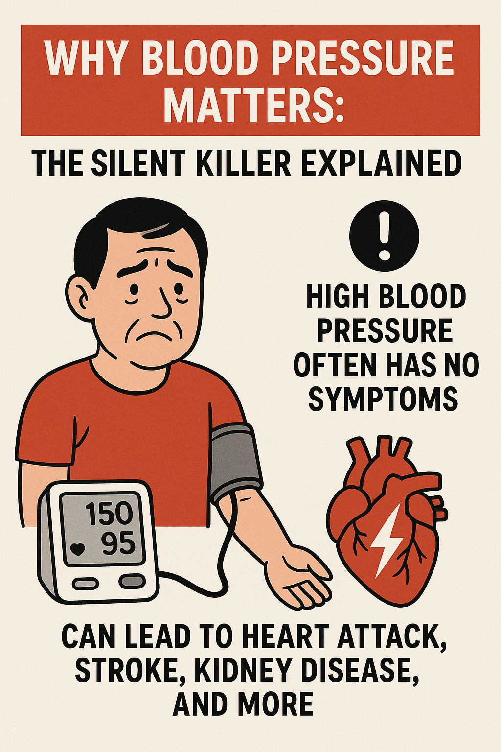
of drugs used in treatment of heart failure, so let s get right into it. Heart failure is simply defined as a chronic,
progressive disorder in which the heart muscle is unable to pump enough blood to meet the
body’s needs. In a normal heart, the upper chambers called
the atria and the lower chambers called the ventricles squeeze and relax in turn to move
blood through the body.
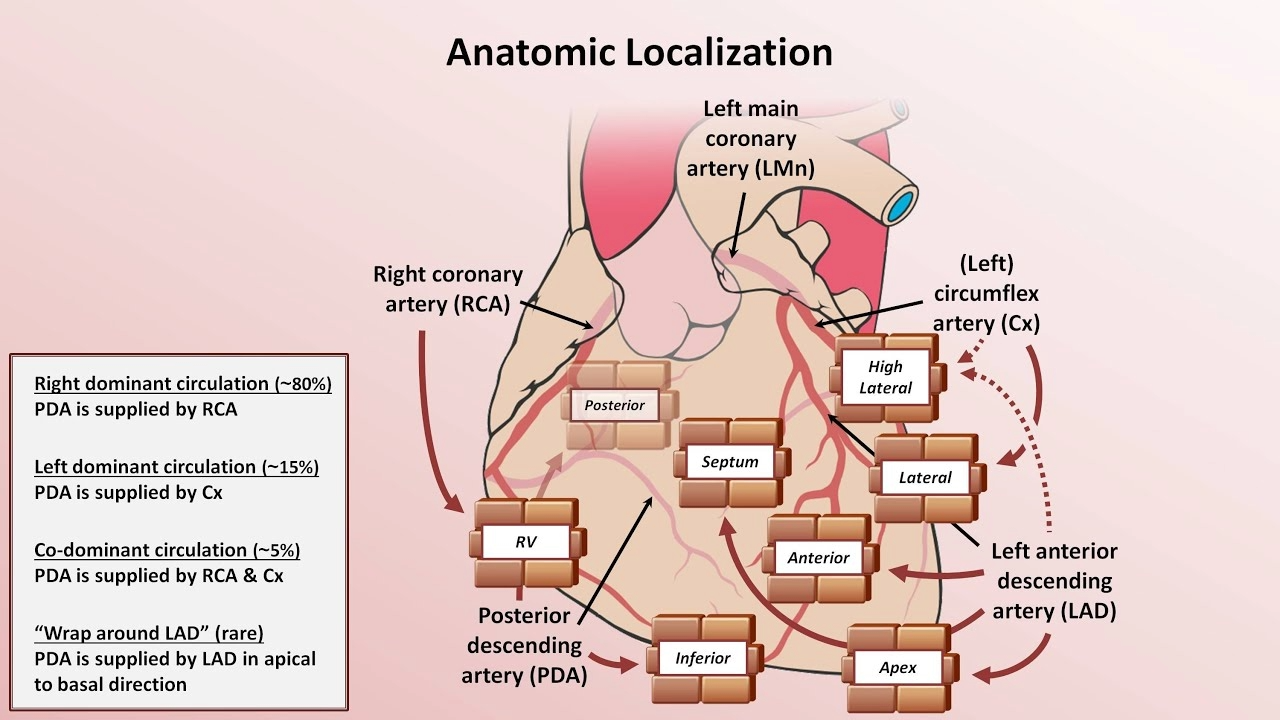
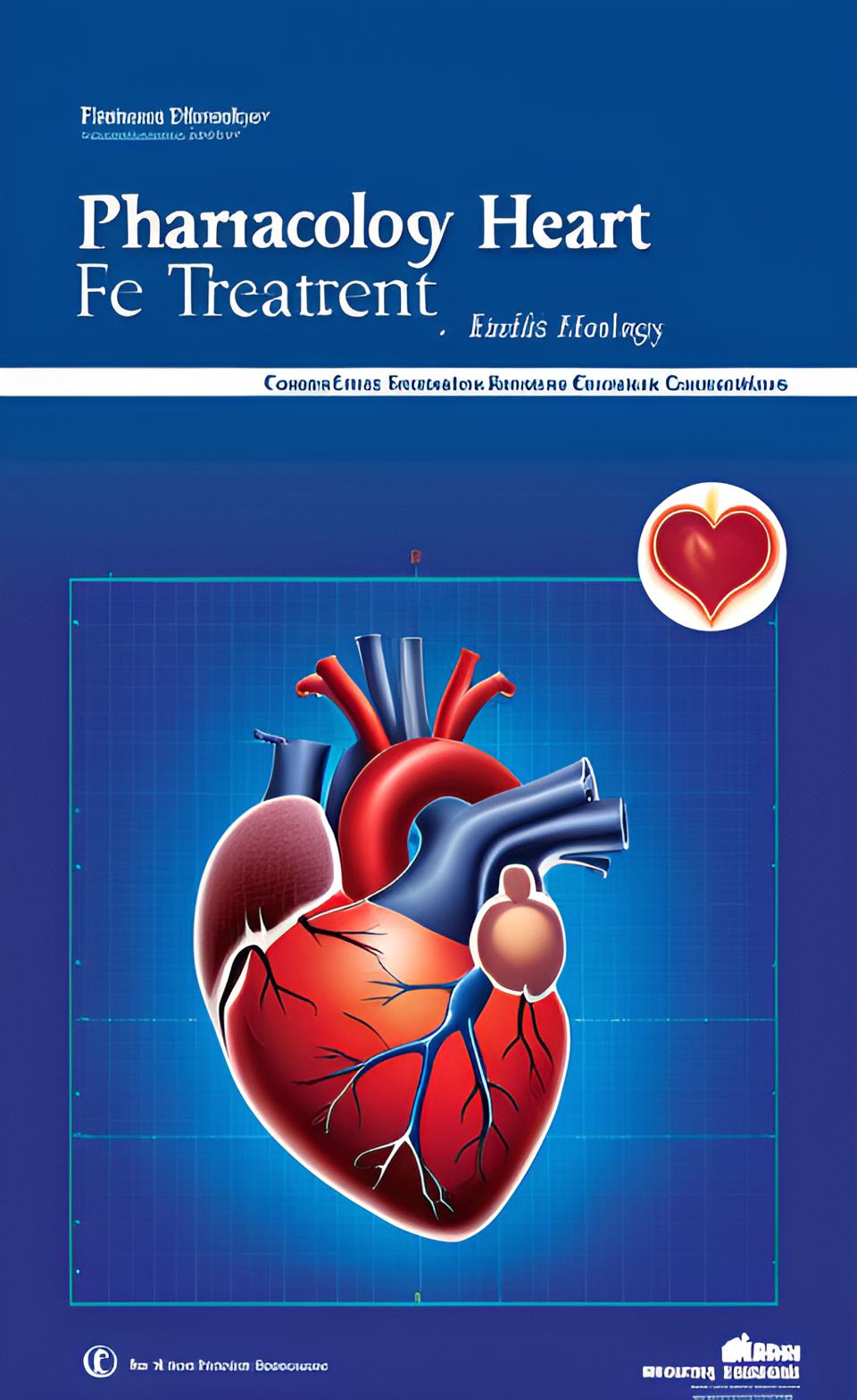
Cardiac Blood Flow: Four Major Steps
Now blood flows through the heart and lungs
in four major steps. First, the oxygen-poor blood that has already
circulated through the body is received by the right atrium, which in turn pumps it over
to the right ventricle. Secondly, the right ventricle pumps the blood
through the pulmonary artery into the lungs, where it picks up oxygen. Thirdly, the pulmonary vein empties oxygen-rich
blood, from the lungs into the left atrium, which in turn pumps it to the left ventricle. And finally the left ventricle pumps oxygenated
blood through the aorta to the rest of the body.
The Frank-Starling Law and Heart Failure
Now, the Frank-Starling law of the heart is
a basic physiological principle that describes how the heart is able to move blood through
the body in a regulated way by pumping out as much blood as it receives. Specifically, this law states that increased
filling of the ventricle results in greater contraction force and thus a rise in the cardiac
output. In heart failure however this mechanism fails,
as the ventricle is loaded with blood to the point where heart muscle contraction becomes
less efficient.
Types of Heart Failure: Systolic vs Diastolic
Now, depending on the primary cause, heart
failure can manifest itself as either systolic or diastolic dysfunction. In systolic heart failure the heart muscle
becomes weak and cannot squeeze as much blood out. Poor ventricular contractility leads to reduction
in the amount of blood pumped out of the ventricle, which we refer to as ejection fraction. While the normal ejection fraction can range
between 50 and 75%, heart failure due to systolic dysfunction is typically associated with an
ejection fraction of less than 40%. For this reason the systolic heart failure
is most commonly referred to as Heart Failure with Reduced Ejection Fraction (HFrEF). On the other hand, in diastolic heart failure
the heart squeezes normally, but becomes stiff and cannot adequately relax to allow for normal
ventricular filling. As a result, patients with diastolic heart
failure have relatively normal ejection fraction although stroke volume and cardiac output
are reduced. Because of this, diastolic failure is most
commonly referred to as Heart Failure with Preserved Ejection Fraction (HFpEF).
Compensatory Mechanisms in Heart Failure
Now, in the presence of heart failure, in
order to counteract the effect of falling cardiac output and thus reduced perfusion
to vital organs, the body will try to compensate via two tightly regulated mechanisms. The first one involves the increase in sympathetic
nervous system activity. In the face of a reduced cardiac output, the
arterial baroreceptors located in the aortic arch and carotid sinus will sense changes
in blood pressure leading to the release of norepinephrine that in turn stimulates beta-1
receptors located in the SA node, myocardium and the ventricular conduction system. Stimulation of these receptors increases heart
rate and cardiac contractility leading to greater stroke volume. Because heart rate and stroke volume are components
of cardiac output, which is simply equal to the product of the two, when they both increase,
cardiac output will also increase to maintain adequate blood pressure and thereby perfusion
to vital organs. Moreover, increased sympathetic nerve traffic
to the kidney also activates ?1-adrenergic receptors located on juxtaglomerular cells
causing them to release an enzyme responsible for regulation of blood pressure and volume
called renin.
Renin-Angiotensin-Aldosterone System (RAAS)
And this brings us to the second major compensatory
mechanism, which involves activation of the renin angiotensin aldosterone system. So, in addition to sympathetic nerves directly
stimulating renin secretion via ?1 receptors, the release of renin from the juxtaglomerular
cells is also regulated by two other primary mechanisms which are; the renal vascular baroreceptors
that stimulate renin secretion in response to low renal perfusion pressure, and the macula
densa cells of the distal nephron that stimulate renin secretion in response to fall in sodium
chloride concentration. Now once released into the blood, renin acts
upon a circulating substrate that is primarily supplied by the liver called angiotensinogen
to produce angiotensin I. On passing through the pulmonary circulation
angiotensin I is converted into angiotensin II by another enzyme, which is abundant in
the lungs called angiotensin-converting enzyme (ACE for short). Now, circulating angiotensin II exerts its
action by binding to various receptors throughout the body with most of its effects being mediated
via angiotensin II type 1 receptor (abbreviated as AT1). These include stimulation of AT1 receptors
in the endothelium of systemic arterioles, which leads to vasoconstriction; stimulation
of angiotensin receptors in the brain, which causes the pituitary to release antidiuretic
hormone (ADH for short), which in turn binds to specific vasopressin II receptors in the
collecting ducts of the nehpron and promotes reabsorption of water back into the circulation;
and finally, angiotensin II also acts on the angiotensin receptors in the adrenal cortex
to stimulate the release of a steroid hormone called aldosterone, which in turn binds to
nuclear mineralocorticoid receptor within the cells of the distal tubule and the collecting
duct where it increases expression of genes that encode epithelial sodium channels and
the sodium/potassium pump (Na/K ATPase) thereby promoting sodium and water reabsorption and
potassium secretion causing increase in plasma volume and blood pressure. Furthermore, vasoconstriction and fluid retention
elevates venous and capillary hydrostatic pressures, forcing additional fluid out of
the blood into the tissue leading to edema particularly in the feet and legs. The increased peripheral resistance and greater
blood volume also place further strain on the heart and accelerate the process of damage
to the myocardium leading to structural cardiac remodeling.
Natriuretic Peptides and Counter-Regulation
At this point, in the final attempt to maintain
circulatory system homeostasis, the body will try to counterbalance overstimulation of the
renin angiotensin aldosterone system (RAAS) and sympathetic nervous system by activating
cardioprotective natriuretic peptides. Specifically, in response to increased myocardial
stretch and volume overload, atria begin to secrete A-type natriuretic peptide (ANP) and
ventricles begin to secrete B-type natriuretic peptide (BNP), and in response to increased
levels of pro-inflammatory mediators resulting from cardiac injury, vascular endothelial
cells begin to secrete C-type natriuretic peptide (CNP). Now the main role of these natriuretic peptides
is to counter the effects of volume overload and adrenergic activation by stimulating sodium
and water excretion, promoting myocardial relaxation, inhibiting cardiac hypertrophy
and fibrosis, suppressing sympathetic outflow, and stimulating vasodilation. However, in the end, even this counter response
is not enough to save the failing heart. As heart failure advances, further activation
of the renin angiotensin aldosterone system and the sympathetic nervous system ultimately
overcomes the short-lived beneficial effects of the natriuretic peptides.
Transition to Treatment
And this brings us to the second part of this
lecture that is the treatment of heart failure. Now the pharmacological management of patients
with heart failure is complex and may require the use of several classes of drugs. So now let s discuss these one by one starting
with beta-blockers.
Beta-Blockers
So beta-blockers work by binding to beta-1
receptors in the heart and subsequently blocking the action of norepinephrine thereby reducing
heart rate and contractility which in turn decreases cardiac output and blood pressure. As a side note here, keep in mind that decreased
heart rate allows more diastolic filling time so the stroke volume is typically not reduced.
Now, similarly via blockade of the ?1 receptors
of the renal juxtaglomerular complex, certain beta-blockers may also reduce renin secretion,
thereby reducing the severity of angiotensin II-induced vasoconstriction as well as aldosterone-induced
volume expansion. It s important to remember that this however
is not beta-blockers primary mechanism of action. Among several beta-blockers on the market,
currently only three have proven to reduce mortality in heart failure patients; these
are Bisoprolol, Carvedilol, and Metoprolol. Out of these three, Carvedilol has a unique
pharmacological property in that it not only blocks beta-1 receptors in the heart but also
alpha-1 receptors located on the smooth muscles of arteries and veins. By preventing norepinephrine from activating
the alpha-1 receptor, Carvedilol causes vessels to dilate thereby reducing total peripheral
resistance.
ACE Inhibitors
All right, moving on to the next class of
drugs for heart failure that is angiotensin-converting enzyme (ACE) inhibitors. Drugs in this class selectively inhibit the
angiotensin-converting enzyme, which in turn reduces angiotensin II production and its
effects on vasoconstriction as well as ADH and aldosterone secretion. In addition to this, inhibition of ACE, increases
levels of a potent vasoactive peptide called bradykinin. Unlike angiotensin II, which is a vasoconstrictor,
bradykinin is an endogenous vasodilator, which is normally degraded by ACE. So when ACE inhibition occurs, while angiotensin
II levels drop, bradykinin levels rise. As a result the blood vessels become dilated,
total peripheral resistance is reduced and blood pressure is lowered thereby reducing
the effort needed to pump blood around the body. Drugs in this class include Captopril, Enalapril,
Fosinopril, Lisinopril, Quinapril and Ramipril.
Angiotensin Receptor Blockers (ARBs)
Another related class of drugs called angiotensin
receptor blockers (ARBs) also works on the same angiotensin pathway. However instead of blocking the enzyme that
drives angiotensin II production, ARBs work by binding to AT1 receptors located on vascular
smooth muscle as well as other tissues such as heart directly blocking the actions of
angiotensin II. As a result, the effects are similar to ACE
inhibitors that is less vasoconstriction and less ADH and aldosterone secretion, which
lowers blood pressure and ultimately prevents damage to the heart and kidneys. Also because ARBs do not inhibit ACE, they
do not cause bradykinin levels to rise. This makes ARBs a good alternative to ACE
inhibitors as more bradykinin not only contributes to the vasodilation but also contributes to
some of the side effects of ACE inhibitors such as cough and angioedema. Drugs in this class include Candesartan, Losartan,
Telmisartan, and Valsartan.
ARNI: Angiotensin Receptor–Neprilysin Inhibitor
Now despite being treated with an ACE inhibitor
or angiotensin receptor blocker many heart failure patients continue to suffer from cardiovascular
events. As a result increasing the beneficial effects
of natriuretic peptides has gained significant interest as a therapeutic approach in the
management of heart failure leading to development of a new class of drugs called angiotensin
receptor-neprilysin inhibitor. Now, neprilysin is a circulating enzyme that
degrades several endogenous vasoactive peptides, including ANP, BNP, and CNP and thus terminates
their positive actions. Angiotensin receptor-neprilysin inhibitor
simply combines angiotensin receptor blocker and neprilysin inhibitor to simultaneously
block angiotensin II receptor as well as inhibit neprilysin enzyme thereby preventing it from
breaking down natriuretic peptides. This results in increased longevity of natriuretic
peptides as well as enhancement of their beneficial effects. The example of drug that belongs to this class
is Sacubitril/Valsartan.
Aldosterone Antagonists (Potassium-Sparing Diuretics)
Now, another shortfall of ACE inhibitors and
angiotensin receptor blockers (ARBs) is that in some cases they don t suppress the excessive
formation of aldosterone sufficiently. Therefore, select patients with moderate to
severe heart failure can also benefit from another class of drugs called aldosterone
antagonists. Aldosterone antagonists work by competitively
blocking the binding of aldosterone to the mineralocorticoid receptor thereby decreasing
the reabsorption of sodium and water as well as decreasing the excretion of potassium leading
to cardioprotective effects. For this reason we also refer to this class
of drugs as Potassium-sparing diuretics. The examples of drugs that belong to this
class are Eplerenone and Spironolactone.
Loop Diuretics for Symptom Relief
Now, although aldosterone antagonists have
been shown to lower blood pressure and exert some diuretic effect, in order to alleviate
symptoms of volume overload, a more potent class of drugs called loop diuretics is needed.
So the primary use of loop diuretics is to
relieve symptoms associated with pulmonary congestion and peripheral edema. Loop diuretics achieve this by inhibiting
the luminal sodium-potassium-chloride cotransporter located in the thick ascending limb of the
loop of Henle where about 20% to 30% of the filtered sodium is managed. As a result, in contrast to other diuretic
agents, loop diuretics reduce reabsorption of a much greater proportion of sodium. This sodium is then excreted, along with the
water that follows it, leading to significant decrease in plasma volume, cardiac workload
and oxygen demand thus relieving signs and symptoms of volume excess. Drugs that belong to this class include Bumetanide,
Furosemide and Torsemide.
Vasodilators: Isosorbide Dinitrate and Hydralazine
Now, in some cases when a patient is truly
intolerant of ACE inhibitors or angiotensin receptor blockers (ARBs), usually because
of significant renal dysfunction, the blood pressure can be controlled with another class
of drugs referred to as vasodilators. There are two drugs in this class that are
typically used in treatment of heart failure. The first one is Isosorbide dinitrate, which
releases nitric oxide (NO) in the vascular smooth muscle cell that subsequently activates
guanylyl cyclase (GC), an enzyme that catalyzes the formation of cyclic guanosine monophosphate
(cGMP) from guanosine triphosphate (GTP). Increased intracellular cGMP in turn activates
a series of reactions that cause decrease in intracellular calcium concentrations. And because calcium drives the contraction
this decrease ultimately leads to smooth muscle relaxation and thus vasodilation. Now, in contrast to isosorbide, the second
drug that is Hydralazine appears to have multiple effects on the vascular smooth muscle, which
include; stimulation of nitric oxide release from the vascular endothelium stimulating
cGMP production and decreasing calcium concentration, opening of potassium channels, and inhibition
of calcium release from the sarcoplasmic reticulum, which altogether contribute to smooth muscle
relaxation and subsequent vasodilation.
Digoxin: Positive Inotropy
Finally, before we end, I wanted to briefly
mention one more drug that can be used in management of heart failure particularly in
patients intolerant to ACE inhibitors or beta-blockers that is Digoxin. Now the mechanism of action of Digoxin is
sort of the opposite of the vasodilators one, in that it is used to increase cells’ contractility,
specifically the contractility of cardiac muscle cells. Digoxin accomplishes that by inhibiting the
sodium potassium ATPase pump in cardiac muscle cells, which is responsible for moving sodium
ions out of the cell and bringing potassium ions into the cell. As a result of this inhibition, when sodium
concentration in the cardiac cell increases, another electrolyte mover known as sodium-calcium
exchanger pushes the excess sodium ions out while bringing additional calcium ions in.
This in turn causes an increase in the intracellular
calcium, which is then available to the contractile proteins. The end result is increased force of contraction
and thus increased cardiac output. And with that I wanted to thank you for watching
I hope you enjoyed this video and as always stay tuned for more.
Would you like me to turn this into a formatted blog document on your Canvas or keep it as-is here?