
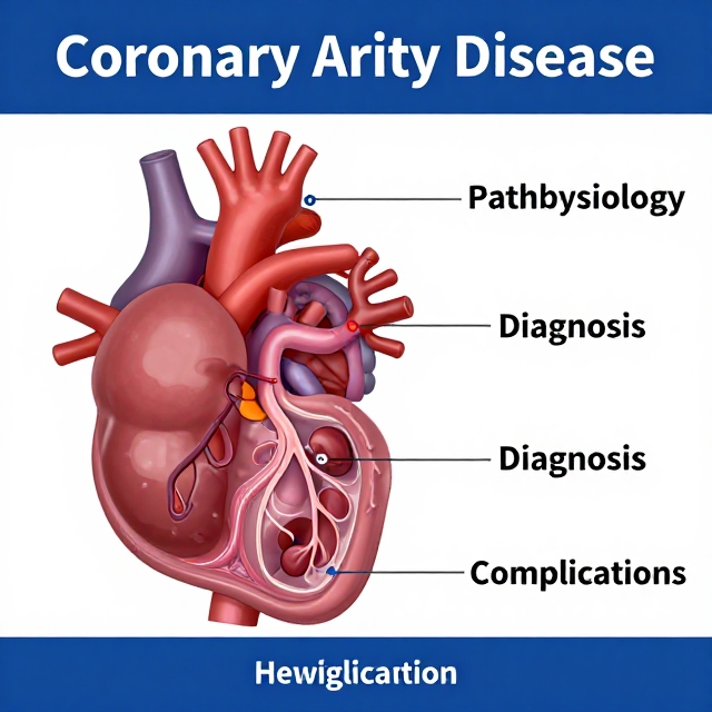
Coronary artery disease (CAD) is one of the most common forms of heart disease. It develops when the arteries that supply blood to the heart muscle become narrowed or blocked, most often because of atherosclerosis.
When coronary arteries cannot deliver enough oxygen-rich blood to the heart muscle, patients may develop angina, shortness of breath, or other symptoms. If a coronary artery becomes severely blocked, it can lead to a heart attack (myocardial infarction).
Understanding the causes, warning signs, diagnostic tests, complications, and treatment options for CAD can help patients recognize potentially serious symptoms and seek medical attention promptly.
What Is Coronary Artery Disease?
Coronary artery disease occurs when plaque builds up inside the coronary arteries. These arteries are responsible for supplying oxygen and nutrients to the myocardium, or heart muscle.
The major coronary vessels include:
- Left anterior descending (LAD) artery: Supplies much of the anterior left ventricle and interventricular septum.
- Left circumflex (LCx) artery: Primarily supplies the lateral portions of the left ventricle.
- Right coronary artery (RCA): Supplies much of the right ventricle and portions of the inferior heart.
- Posterior descending artery (PDA): Supplies portions of the posterior/inferior heart and is commonly a branch of the RCA in right-dominant circulation.
The clinical consequences of CAD depend on how severely an artery is narrowed or blocked and whether the obstruction develops gradually or suddenly.
What Causes Coronary Artery Disease?
The most common underlying cause of CAD is atherosclerosis.
Atherosclerosis occurs when cholesterol, inflammatory cells, calcium, and other substances accumulate within the arterial wall and form an atherosclerotic plaque.
Over time, a plaque can:
- Narrow the coronary artery.
- Reduce blood flow to the myocardium.
- Limit oxygen delivery during increased physical activity.
- Rupture and trigger formation of a blood clot.
- Cause partial or complete coronary artery obstruction.
A stable plaque can produce predictable exertional angina, while plaque rupture can result in an acute coronary syndrome.
Major Risk Factors for CAD
Several factors increase the risk of developing coronary artery disease.
Smoking
Smoking damages the vascular endothelium, promotes inflammation, increases thrombosis risk, and accelerates atherosclerosis.
High Cholesterol
Abnormal lipid levels, particularly elevated LDL cholesterol, contribute significantly to plaque formation. Low HDL cholesterol can also be associated with increased cardiovascular risk.
High Blood Pressure
Hypertension increases stress on arterial walls and contributes to endothelial injury and atherosclerosis.
Diabetes
Diabetes is an important cardiovascular risk factor because chronically elevated blood glucose contributes to vascular damage and accelerates atherosclerosis.
Age
The risk of coronary artery disease increases with age.
Family History
A family history of premature coronary artery disease can increase an individual’s cardiovascular risk.
Other important risk factors include obesity, physical inactivity, unhealthy dietary patterns, and certain inflammatory or metabolic conditions.
Stable Coronary Artery Disease and Angina
In stable CAD, an atherosclerotic plaque gradually narrows a coronary artery. Blood flow may be adequate when the person is resting but insufficient when the heart requires more oxygen.
Physical activity increases:
- Heart rate
- Blood pressure
- Myocardial workload
- Oxygen demand
If the coronary arteries cannot increase blood flow sufficiently, myocardial ischemia can occur.
Typical Stable Angina
Stable angina commonly:
- Occurs with physical exertion or emotional stress
- Feels like pressure, squeezing, heaviness, or tightness in the chest
- Improves with rest
- May improve with prescribed nitroglycerin
Pain may radiate to the arm, shoulder, neck, jaw, or back.
Acute Coronary Syndrome
Acute coronary syndrome (ACS) occurs when there is sudden reduction in coronary blood flow, commonly because an atherosclerotic plaque ruptures and a thrombus forms.
The major clinical categories are:
- Unstable angina
- NSTEMI
- STEMI
Unstable Angina
Unstable angina involves myocardial ischemia without detectable myocardial necrosis.
Symptoms may:
- Occur at rest
- Become more frequent
- Become more severe
- Last longer than previous episodes
Cardiac troponin levels are not elevated because significant myocardial necrosis has not occurred.
NSTEMI
A non-ST-elevation myocardial infarction (NSTEMI) occurs when myocardial injury and necrosis occur without the classic persistent ST-segment elevation seen in STEMI.
Patients generally have elevated cardiac troponin levels.
The ECG may demonstrate:
- ST-segment depression
- T-wave inversion
- Other ischemic changes
STEMI
A ST-elevation myocardial infarction (STEMI) is generally associated with acute coronary artery occlusion and myocardial injury producing characteristic ST-segment elevation.
STEMI is a medical emergency requiring rapid reperfusion when appropriate.
Common Symptoms of CAD and Heart Attack
Typical symptoms include:
- Chest pressure or squeezing
- Chest tightness
- Pain or discomfort with exertion
- Shortness of breath
- Sweating
- Nausea
- Fatigue
- Pain spreading to the arm, shoulder, jaw, neck, or back
Some patients, particularly older adults and people with diabetes, may have atypical symptoms or an anginal equivalent, such as unexplained shortness of breath, nausea, fatigue, or upper abdominal discomfort.
Severe or new chest discomfort, especially when accompanied by shortness of breath, sweating, fainting, or nausea, requires urgent medical evaluation.
How Is Coronary Artery Disease Diagnosed?
The diagnostic approach depends on the patient’s symptoms and whether an acute coronary syndrome is suspected.
Electrocardiogram (ECG/EKG)
An ECG is one of the first tests used when acute coronary syndrome is suspected.
Possible findings include:
- ST-segment elevation
- ST-segment depression
- T-wave inversion
- Pathologic Q waves
- Rhythm abnormalities
Cardiac Troponin
Cardiac troponin is a major biomarker used to identify myocardial injury.
A rise or fall in troponin, interpreted in the appropriate clinical context, supports the diagnosis of myocardial infarction.
Echocardiogram
An echocardiogram can identify abnormalities in heart-wall movement and assess ventricular function.
Wall-motion abnormalities can sometimes help correlate myocardial injury with a particular coronary territory.
Stress Testing
For selected patients with suspected stable CAD, stress testing can evaluate whether exercise or pharmacologic stress produces evidence of myocardial ischemia.
Possible approaches include:
- Exercise ECG
- Stress echocardiography
- Myocardial perfusion imaging
- Pharmacologic stress testing
The appropriate test depends on the patient’s ability to exercise and their baseline ECG and clinical characteristics.
Coronary CT Angiography
Coronary CT angiography (CCTA) is a noninvasive imaging test that can visualize coronary arteries and detect plaque and narrowing in appropriate patients.
Coronary Angiography
Invasive coronary angiography provides detailed visualization of the coronary arteries and can identify significant obstructive lesions.
It also allows clinicians to proceed directly to certain forms of revascularization when indicated.
Localizing a STEMI on ECG
Different ECG leads correspond to different regions of the heart.
| Infarct location | Common ECG leads | Commonly associated artery |
|---|---|---|
| Anterior | V1–V4 | LAD |
| Inferior | II, III, aVF | Usually RCA |
| Lateral | I, aVL, V5–V6 | LCx or other lateral supply |
| Posterior | V7–V9 | PDA/posterior circulation |
These relationships are useful clinically, although coronary anatomy varies between individuals.
Patients with an inferior myocardial infarction may also require right-sided ECG leads to evaluate for right ventricular involvement.
Complications of Myocardial Infarction
A myocardial infarction can damage cardiac muscle and produce serious complications.
Arrhythmias
Arrhythmias can occur early after myocardial infarction.
Depending on the affected coronary territory, patients may develop:
- Bradycardia
- AV block
- Ventricular tachycardia
- Ventricular fibrillation
Ventricular fibrillation can result in sudden cardiac arrest.
Acute Heart Failure
A large myocardial infarction can significantly reduce ventricular contractility.
Reduced left ventricular function can cause:
Myocardial injury → reduced contractility → reduced cardiac output → hypotension → possible cardiogenic shock
Blood backing up into the pulmonary circulation can also produce pulmonary edema and shortness of breath.
Cardiogenic Shock
Severe myocardial damage can cause the heart to become unable to provide adequate circulation to vital organs.
Possible findings include:
- Severe hypotension
- Cold extremities
- Altered mental status
- Reduced urine output
- Pulmonary congestion
- Evidence of poor organ perfusion
Cardiogenic shock is a life-threatening complication requiring immediate treatment.
Pericarditis After Myocardial Infarction
Inflammation can extend from damaged myocardial tissue to the surrounding pericardium.
Pericarditis typically causes:
- Sharp or pleuritic chest pain
- Pain that may improve when leaning forward
- Pain that worsens with inspiration
- Pericardial friction rub
- Sometimes fever
Post-infarction pericarditis can occur relatively early, while Dressler syndrome is a later immune-mediated post-myocardial-infarction syndrome.
Mechanical Complications
Although less common in the modern reperfusion era, mechanical complications of myocardial infarction can be extremely serious.
Ventricular Septal Rupture
Necrotic ventricular septal tissue can rupture, creating a ventricular septal defect.
A new harsh holosystolic murmur after an MI should raise concern for this complication.
Papillary Muscle Rupture
Papillary muscle rupture can cause acute severe mitral regurgitation.
It is classically associated with an inferior myocardial infarction involving the RCA territory.
Possible findings include:
- Sudden pulmonary edema
- Hypotension
- New holosystolic murmur
- Acute heart failure
Left Ventricular Free-Wall Rupture
Rupture of the ventricular free wall can cause blood to enter the pericardial space.
This may result in:
Hemopericardium → cardiac tamponade → obstructed cardiac filling → severe hemodynamic compromise
Possible findings of cardiac tamponade include hypotension, elevated jugular venous pressure, and muffled heart sounds.
Left Ventricular Pseudoaneurysm
A contained ventricular rupture can form a pseudoaneurysm. Blood can stagnate within the abnormal cavity, increasing the risk of thrombus formation and systemic embolization.
Treatment of Coronary Artery Disease
Treatment depends on whether the patient has stable CAD, unstable angina, NSTEMI, or STEMI.
Lifestyle Changes
Important measures include:
- Stopping smoking
- Maintaining a healthy weight
- Regular physical activity when medically appropriate
- Following a heart-healthy dietary pattern
- Controlling blood pressure
- Managing diabetes
- Controlling cholesterol
- Following prescribed medications
Medications
Depending on the patient’s condition, treatment may include:
- Antiplatelet medications
- Statins
- Beta blockers
- Nitroglycerin
- Other anti-anginal medications
- ACE inhibitors or ARBs in appropriate patients
- Other cardiovascular medications based on individual risk and comorbidities
Medication selection should be individualized by a qualified healthcare professional.
Percutaneous Coronary Intervention (PCI)
PCI is a minimally invasive procedure used to restore blood flow through a narrowed or blocked coronary artery.
During PCI, a catheter is advanced into the coronary artery. A balloon may be used to open the narrowed area, followed by placement of a coronary stent when appropriate.
After stent placement, antiplatelet therapy is commonly required to reduce the risk of stent thrombosis. The specific medication combination and duration depend on the clinical situation and bleeding risk.
Coronary Artery Bypass Grafting (CABG)
CABG is a surgical procedure that creates an alternative route for blood to flow around blocked coronary arteries.
Grafts may be obtained from vessels such as:
- Internal mammary arteries
- Saphenous veins
- Other suitable arterial conduits
CABG may be preferred in certain patients with complex or extensive coronary disease, including selected patients with left-main disease, multivessel disease, or specific patterns of coronary disease combined with reduced ventricular function.
The choice between PCI and CABG should be determined by the patient’s anatomy, heart function, comorbidities, surgical risk, and recommendations from the treating heart team.
PCI vs. CABG
| Treatment | General concept |
|---|---|
| PCI | Catheter-based treatment using balloon angioplasty and often a stent |
| CABG | Surgery that creates bypass routes around blocked arteries |
Neither treatment is automatically best for every patient. The appropriate approach depends on the severity and location of coronary disease and the patient’s overall health.
How to Reduce Your Risk of CAD
You can reduce cardiovascular risk by:
- Avoiding tobacco.
- Managing blood pressure.
- Keeping LDL cholesterol under appropriate control.
- Managing diabetes.
- Maintaining a healthy weight.
- Exercising regularly as medically appropriate.
- Eating a heart-healthy diet.
- Taking prescribed medications consistently.
- Attending recommended cardiovascular checkups.
- Discussing your personal cardiovascular risk with a healthcare professional.
When to Seek Emergency Care
Do not ignore sudden or severe chest discomfort.
Seek emergency medical attention if chest pain or pressure is:
- New or severe
- Occurring at rest
- Lasting several minutes
- Repeated or worsening
- Associated with shortness of breath
- Associated with sweating or nausea
- Associated with fainting or severe weakness
- Spreading to the arm, jaw, neck, shoulder, or back
A heart attack can be life-threatening, and early treatment can significantly affect outcomes.
Frequently Asked Questions
What is the most common cause of coronary artery disease?
Atherosclerosis is the most common underlying cause. It involves plaque accumulation within the coronary arteries.
What is the difference between stable angina and unstable angina?
Stable angina typically occurs predictably with exertion and improves with rest. Unstable angina is less predictable and may occur at rest or become more frequent or severe.
What is the difference between NSTEMI and STEMI?
Both are myocardial infarctions. NSTEMI generally does not produce the characteristic ST-segment elevation of STEMI, while STEMI is associated with ST-segment elevation and often an acutely occluded coronary artery.
Can coronary artery disease be treated?
Yes. Treatment can include lifestyle changes, medications, PCI with stenting, CABG, or combinations of these approaches depending on the patient’s condition.
Is coronary artery disease preventable?
Many major cardiovascular risk factors are modifiable. Avoiding smoking, controlling cholesterol and blood pressure, managing diabetes, exercising, maintaining a healthy weight, and following a heart-healthy lifestyle can substantially reduce cardiovascular risk.
Conclusion
Coronary artery disease develops when the coronary arteries become narrowed or blocked, most commonly because of atherosclerosis. It can range from stable exertional angina to life-threatening acute coronary syndromes such as NSTEMI and STEMI.
Recognizing symptoms, understanding cardiovascular risk factors, and obtaining appropriate medical evaluation are essential. Modern treatments—including medications, PCI, and CABG—can restore or improve blood flow and reduce the risk of serious cardiovascular complications.
Medical Disclaimer: This article is intended for educational purposes only and does not replace professional medical advice, diagnosis, or treatment. Anyone experiencing symptoms suggestive of a heart attack should seek emergency medical care immediately.